
| Glaucoma: An Eye Emergency
Race Foster, DVM Marty Smith, DVM Holly Frisby, DVM Drs. Foster & Smith, Inc.
When
most pet owners think of medical emergencies relating to their pet, they
picture incidents like the
What is glaucoma? Glaucoma is a very common disease in humans and also very common in dogs. Some medical conditions are difficult to understand, but glaucoma is not. It merely means that the pressure fluid inside the eyeball (or globe) is excessively high. When this occurs, internal structures are destroyed. Its similar to high blood pressure causing a vessel within the brain to rupture and blood flooding into the surrounding tissue crushing brain cells. In lay terms this is referred to as a stroke. In the eye, the elevation of the pressure of the internal fluid to dangerous levels affects almost every tissue inside the globe. In most cases, this renders the eye blind and useless. 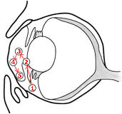 In glaucoma, elevations in the pressure of the aqueous humor are most frequently caused by this fluid not being able to drain correctly from the eye. Additional fluid is constantly being produced and if an equal amount does not leave the globe, then the pressure starts to rise. It is very similar to a water balloon. As more water is added, the balloon stretches more and more. In the case of the balloon, it finally ruptures. The eyeball is strong enough that it does not stretch to any great degree so the force of the increasing pressure is felt by the eyes internal structures. They are either crushed or displaced. In either case, they are rendered nonfunctional. Types of glaucoma We refer to glaucoma as being primary or secondary. Primary Glaucoma: Primary glaucoma occurs in an animal because it possesses physical or physiologic traits that predispose it to glaucoma. This is usually predetermined by genetics. For instance, eyes may have drainage pores that are too small or naturally narrow angles such that the fluid has a difficult time making its way out of the globe. Primary glaucoma is most common seen in Beagles, Cocker Spaniels, and Basset Hounds. It also occurs in Norwegian Elkhounds, Miniature Poodles, Dachshunds, Bouvier des Flanders, and a few others. The exact cause of primary glaucoma is slightly different in each of these breeds, but suffice to say that the end result is the same. In these breeds, even though the animal carries the trait for this disorder, the disease itself does not usually develop until the dog is two or three years of age or older. It appears that developmental changes continue to occur until the animal is mature and then an additional period of time is necessary for clinical signs to appear. With primary glaucoma, both eyes are rarely effected equally or at the same time. The disease usually occurs in one eye months or even years before it affects the second one. Secondary Glaucoma: Secondary glaucoma means that the disease is secondary to, or caused by, another condition. A common example is a penetrating wound to the eye. This often causes an inflammation and the fluid may become too thick to flow out through the drainage pores or it might cause scar tissue to form with the drainage angle, itself, closing. A brief list of the causes of secondary glaucoma would include bleeding in the eye, inflammation within the eye, luxation or displacement of the lens, attachments or scarring between the iris and the lens, degeneration of the structure within the drainage angle, or anything that causes the angle to narrow or close. With inflammation or bleeding into the eye, glaucoma is simply caused by the drainage pores or angle becoming so clogged that adequate fluid cannot escape the eye. Luxation of the lens means that the small attachments holding it in place have weakened or broken down and the lens has moved forward. When this happens, it rests against the iris and blocks the opening (or pupil). An attachment or growing together of the lens and the iris can happen with or without luxation but the result is the same - the pupil is closed and the fluid cannot pass through. The cells and tissue of the drainage angle can degenerate or wear out with age, causing them to lose their ability to function correctly. In these cases, the cells sometimes change type or scar, preventing drainage. As can be imagined, a narrowing of the drainage angle can be caused by many different events. If the base of the iris or ciliary body becomes swollen or enlarged, the iris and cornea are pushed together and the fluid will be unable to make it to the deeper reaches of the angle. Drainage will be impossible. This can be caused by tumors, infections, inflammations, etc. Signs of glaucoma All of this information will hopefully give you an understanding of why or how glaucoma occurs. What is even more important for you, the owner, is to be able to recognize the earliest signs of glaucoma. Just so there is no misunderstanding, if treatment for glaucoma in the dog is not started within a few days or in some cases a few hours of the pressure increase, vision will probably be lost completely from the affected eye. The pressure can crush the cells of the retina and optic nerve, rendering them nonfunctional. It can break down the structures holding the lens in place and it can cause damage to the iris and cornea. After these internal changes have occurred, the eyeball itself swells in size, tilts off to the side, and all the surface blood vessels enlarge giving it the appearance of a large, ugly, bruised radish. The early signs of glaucoma that an owner may watch for or notice are pain, a dilated pupil, cloudiness within the cornea and/or an increase in the size of the blood vessels in the white portion of the eye. You may notice that one eye seems larger or protrudes more than the other. Most animals will not display all of these signs initially, maybe only one or two. The pain may be indicated by the dog rubbing his eye with his paw, against the furniture or carpet, or your leg. This is a common, and often unnoticed, early sign. Some dogs will also seem to flutter the lids or squint with one eye. The pupil of the affected eye will usually dilate early in the course of the condition. It may still react to all bright light shining in it, but it will do so very slowly. Remember that glaucoma, even primary glaucoma, is usually going to initially affect just one of the eyes. If the pupil in one eye is larger than in the other, something is definitely wrong and it may be glaucoma. The cornea is normally perfectly clear, you cannot really see it. Its main function is to hold the liquid portion of the eye in place but at the same time allow light to easily pass through. Glaucoma causes it to lose this clearness and become cloudy. This can occur with other disorders and all deserve medical attention. With glaucoma, the elevated pressure stretches the cornea and tears apart the small protein fibers that give it strength. This change in its internal structure is what causes the initial cloudy appearance. Later in the course of the condition, fluid (or edema) enters the layers of the cornea along with tiny blood vessels. Early in some glaucoma cases, the vessels on the white portion of the eye (the sclera), enlarge and increase in number. This would be similar to "bloodshot eyes" in a person, only much worse. Usually, when this occurs major changes have already taken place inside the eyeball and vision may have been lost. If you observe any of these signs, take your dog to the veterinarian for a physical exam. This does not mean next Tuesday, it means immediately - that same day. In many cases, your veterinarian will initiate treatment immediately but may also refer you to a veterinary ophthalmologist who spends at least eight hours of every day working with the eyes of animals. This is important because every case of glaucoma is different and only with a lot of experience can the correct decisions be made. If we delay at all, or an incorrect therapy is chosen, the animal will probably go blind. We may not be able to prevent blindness from occurring, but we can learn from the first eye so that preventative measures can be taken to control or prevent the condition in the second eye. This is especially important in primary glaucoma. Treatment The treatment for glaucoma depends on its cause and severity. There are three goals: Reduce the pressure within the eye
Medical treatment of glaucoma in dogs does not work as well as it does in people. In most cases the only long-term control of glaucoma is achieved through surgery. Various procedures including cyclocryotherapy (liquid nitrogen) can be used depending on the underlying cause of the glaucoma. If neither medical or cyclocryotherapy treatments are effective, there are two options. If there is no pain, infection or neoplasia present, an intraocular prosthesis can be inserted. In this procedure the internal contents of the eye are removed and replaced with a silicone ball. There is generally a high rate of success with this procedure. If all other treatments have failed or the glaucoma is a result of neoplasia or intractable infection, the eye may need to be removed (enucleated). In cases of secondary glaucoma, treatment of the underlying cause is also necessary. This may include antibiotics for bacterial infection, or surgery for the removal of a tumor. Don't Delay Glaucoma is an emergency! If your dog is
showing ocular (eye) pain, a dilated pupil, a cloudy cornea and/or a bloodshot
eye -- get your dog into a veterinary clinic immediately! Whether you want
to believe it or not, it probably is an emergency.
© 2001 Drs. Foster & Smith, Inc.
|